Description:
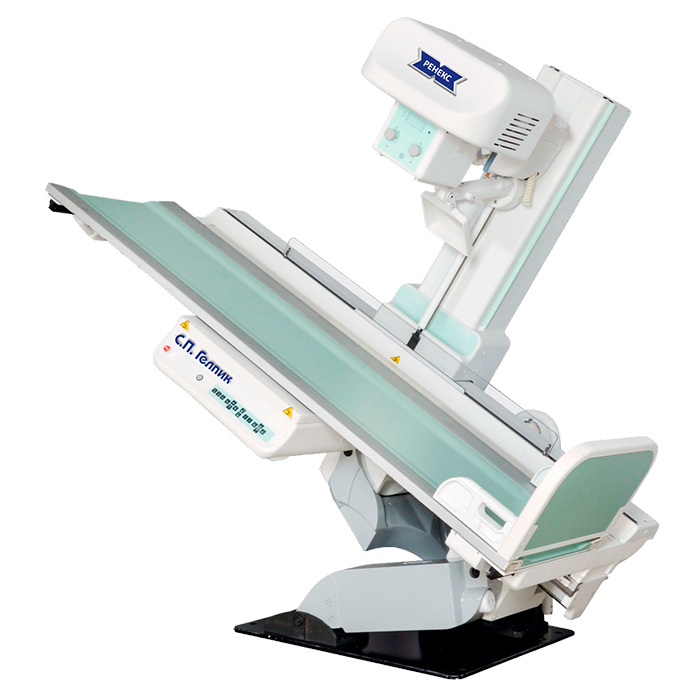
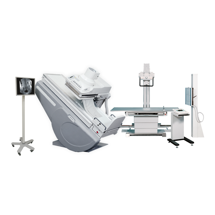
The complex is equipped with an X-ray image amplifier with a high-resolution digital camera for digital fluoroscopy, while radiography can be performed using a digital system based on a flat-panel detector (DR) with one or two flat-panel detectors, or with a digital radiography device (CR) or analog tape cassettes.
Depending on the request, the complex can be equipped with a linear X-ray tomography function, an “auto-tracking” function – automatic tracking and centering of the receiver after the movement of the emitter.
A wide range of selection of complete sets and additional equipment allows you to choose the best option at an optimal cost.
Features and Benefits:
- Routine diagnostics in the digestive tract, chest organs, and the skeleton and skull. Special studies in gynecology, urology and other fields;
- Fluoroscopy with an X-ray image intensifier equipped with a high-resolution digital camera with the last frame hold;
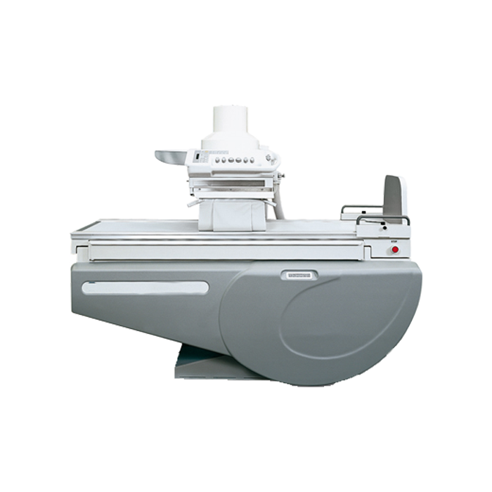
- The image table is equipped with an elevator that allows you to change the height of the deck in the range from 50cm to 90cm from the floor, which allows you to adjust the deck to the height of the gurney;
- High-frequency X-ray power supply with automatic exposure mode, microprocessor control system and self-diagnostics;
- Automatic exposure selection system: two-point, one-point and anatomical programming (organ-automatic);
- The choice of conditions for radiography (tomography) in the organ-automatic mode includes more than 2000 anatomical programs. Possibility of manual correction of organ-automation modes. The set includes a visual atlas of patient positions;
- After each shot, the effective equivalent dose of the patient (in μSv) is displayed;
- Touch interactive control panel. The design of the console with the use of modern technology touch screen allows you to control the operation of the device and take pictures by several touches, which greatly simplifies the work of the X-ray technician;
Digital technology makes it possible to obtain a high-quality diagnostic image on a computer screen instantly during the examination process, which allows you to make express diagnostics. This is extremely important in the admission department of hospitals, in trauma departments, in intensive care units, etc. The use of digital technology in stationary diagnostic devices allows you to fully realize its advantages over analog research methods:
- Lack of an intermediate photoprocess, and as a result – express diagnostics;
- Significant savings in expensive silver-containing photographic materials;
- Practical absence of marriage when taking pictures;
- Significant improvement in the quality of diagnostics due to digital processing of the X-ray image on the computer screen;
- Digital archiving of images on electronic media saves storage space and makes working with the archive easily accessible;
- Sending images electronically;
- Obtaining a hard copy of an X-ray image both on a conventional laser printer with printing on paper, and on other devices with high-quality printing on a transparent base. virtually no rejects when taking pictures, due to the large dynamic range (low sensitivity to exposure errors).
Techical characteristics
Techical characteristics:
System for radiography and tomography Optima Millennium and Moviplan IC:
High-frequency X-ray feeding device “Renex”:
- Supply voltage three-phase 380V +/- 10%;
- Exposure selection system: two-point, one-point and anatomical programming;
- Organ-automatics over 1800 programs. Possibility of manual correction of organ-automation modes;
- Power 65kW;
- The operating frequency of the inverter is 55kHz. Ripple frequency up to 333kHz;
- Anode voltage range for X-ray imaging is 40-150kV;
- Anode voltage step, 1kV or in accordance with the standard series;
- X-ray tube current range for fluoroscopy is 0.5-10mA;
- X-ray tube current range for radiography is 10-800mA (possibly up to 1000mA);
- Electricity amount range is 1-1000mAs;
- The minimum exposure time is 1 ms;
- Indication of the radiation dose in μSv (in the automatic organ mode);
- X-ray mode auto-calibration system;
- Flexible multiprocessor system of microprocessor control system. The ability to adapt to customer requirements;
- The language for displaying parameters and marking controls is Russian.
Equipment:
- Control unit with high voltage module;
- Control panel based on a touch screen 15″, with a remote exposure button;
- High voltage cables 12m with tips (2 pcs).
X-ray tube emitters:
- X-ray emitter Optilix 154/30/50R (Anode angle – 16°, 9000rpm, maximum operating voltage 125kV, focal spots 0.6×0.6mm (30kW) and 1.0×1.0mm (50kW), heat capacity not less than 340kHU), or Analog.
- X-ray emitter Toshiba 7252X (Anode angle – 12°, 9000rpm, maximum operating voltage 150kV, focal spots 0.6×0.6mm (25kW) and 1.2×1.2mm (75kW), heat capacity not less than 300kHU), or Analog.
Digital system for registration of X-ray images based on stationary flat-panel detectors ViVIX-S:
ViVIX-S is a stationary flat panel system for digital radiography. This system is designed for use as part of X-ray diagnostic complexes, in order to accelerate the process of obtaining diagnostic images. The system is a modern alternative to digitizers (CR) and X-ray cassettes with film, eliminating intermediate photoprocesses.
Replaces digitizers and X-ray film cassettes.
The ability to receive digital images and their subsequent processing, zooming, panning images, and other functions that allow operators to see the features of an X-ray image that are difficult to see when using conventional film.
Delivery set
Delivery set:
- Complexes of the “Renex” series for 3 workplaces;
- Stationary flat-panel detector for vertical rack 43x43cm – 2 pc.
- Digital medical imaging device;
- High-frequency power supply with a power of 65kW;
- Dual focus X-ray emitter;
- Workstation of a laboratory assistant with a monitor, LCD, not less than 21 ″;
- Workstation of a doctor with a monitor of a doctor, LCD, at least 24 ″;
- Remote Control;
- Office printer for printing descriptions;
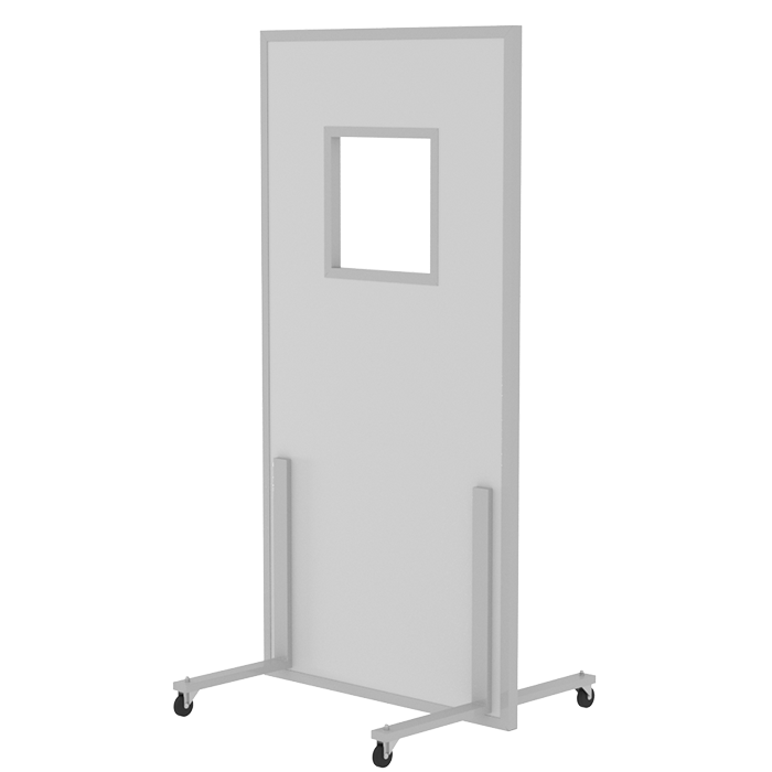
- A set of radiation protection equipment;
- Intercom;
- Dosimeter;
The delivery includes: installation, commissioning, training of hospital personnel by the supplier’s specialists;
Warranty period: 18 months;
Delivery time: within 90 days;
Time of putting the device into operation: within 20 days after delivery, given that subject to the readiness of the premises for the start of installation work and the availability of an approved technological design.
To install the device, a treatment area of 24sq.m is required (SanPiN 2.6.1.1192-03)


