Description:
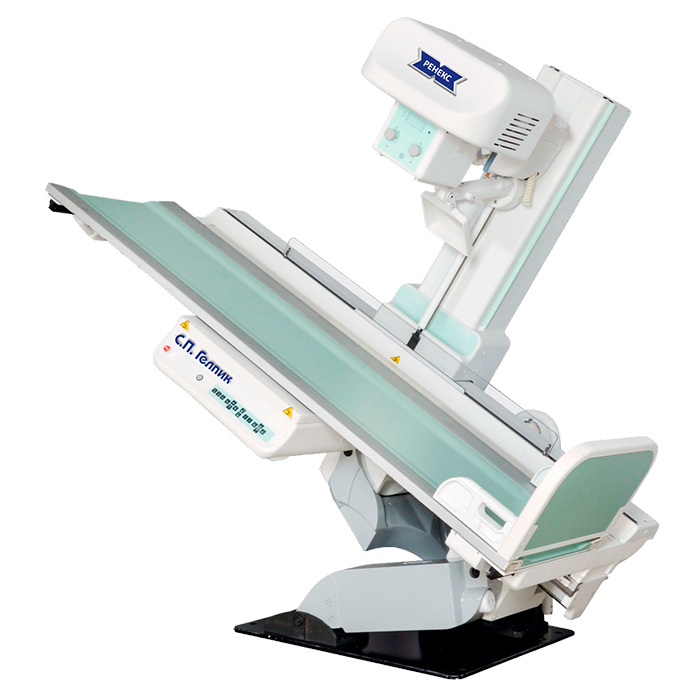
The complex is equipped with a digital dynamic flat-panel detector of 43×43 cm format for radiography, linear tomography and fluoroscopy.
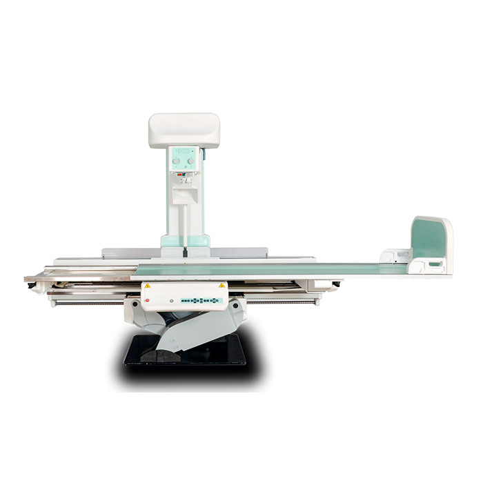
Possibility of fluoroscopy, radiography, tomography at one workplace and at a time, with a vertical, horizontal or tilted position of the patient. One remote-controlled tripod provides all the capabilities of the device for 3 workplaces;
Depending on the version, the complex can be equipped with the function of vertical lifting of the table deck, longitudinal and transverse movement of the table deck, an additional vertical stand, various layouts of screening grids and compression devices.
A wide range of selection of complete sets and additional equipment allows you to choose the best option that takes into account all the needs of the client at an optimal cost.
Features and Benefits:
- KRDC Т20/Т2000 has the most modern functions, such as:
-Function Stitching (Stitching) – obtaining a single image from several, by software stitching. The function is indispensable in traumatology for obtaining a single image of the lower extremities and the spine.
-Digital subtraction angiography is the most informative and accurate method of contrast X-ray examination of blood vessels, which allows you to study in detail the functional state of blood vessels, blood flow, to identify severity and the length of the lesion. Subsequent computer processing allows you to obtain high quality images.
-Tomosynthesis is a method based on performing a series of low-dose images and changing the position in the opposite direction of the receiving device – a flat-panel dynamic detector. Allows you to obtain a reconstructed three-dimensional image of the studied organ.
-The multi-energy study function (dual energy) is a function that allows you to obtain a reconstructed image with suppression of bone or soft tissue, thanks to the conduct of two low-dose exposures with different energies. The main application of this method is chest screening for the diagnosis of neoplasms in the lungs, which, during a routine examination, can be hidden by bone tissue and go unnoticed.
- All functions of the device are controlled from the control room.
- The highest quality of the resulting digital image.
- The program for improving the quality of the image allows you to clearly see the details of the image of both soft tissues and bone structures; Automatic exposure selection system: two-point, one-point and anatomical programming (organ-automatic);
Techical characteristics
Techical characteristics:
Power supply:
- Power: 50/65/80kW
- Anode voltage range for X-ray imaging is 40-150 kV;
- The range of anode voltage variation during fluoroscopy is 40-125 kV;
- X-ray tube current range for radiography is 10-1000 mA;
- X-ray tube current range for fluoroscopy is 0.5-10 mA
- Electricity quantity range is 0.1 – 1000 mAs;
- The minimum exposure time is 0.001 sec;
Telecontrolled tripod:
- Table tilt from +90°/-30 to +90°/-90° with automatic stop in horizontal position;
- Simultaneous movements: table tilt, rack longitudinal movement, table surface movement, rack tilt;
- Table deck size up to 240 x 80 cm;
- Table deck height:
– no more than 86 cm from the floor;
– with a range of motion from 76 cm to 100 cm from the floor;
– with a range of motion from 50 cm to 100 cm from the floor.
- Focus-film distance continuously variable from 115 cm to 180 cm;
- Longitudinal movement of the tripod together with the ESA;
- The speed of the longitudinal movement of the tripod is up to 25 cm/s;
- Three-pole ionization chamber with preamplifier for X-ray exposure meter;
- Linear tomography in both directions;
- Choice of tomography layer height from 0 to 330 mm;
- Automatic collimator;
- Palpation device with automatic movement to the parking area;
Dynamic flat panel detector:
- Scintillator type – Cesium-Iodine (CsI);
- Active detection area is 430×430 mm;
- Resolution capacity is not less than 4.0 lp/mm;
- Detector capacity is not less than 16 bits;
- The size of the matrix of the resulting image is at least 3072×3072 pixels;
- Pixel size is no more than 139 microns;
- Quantum efficiency ratio (DQE) – 75%;
- Full-format image output time – no more than 3 seconds;
- Support for linear tomography mode.
Комплект поставки
Delivery set:
Package included:
- X-ray diagnostic digital complex with a rotary table-tripod KRDC-T20/T2000 – “Renex”;
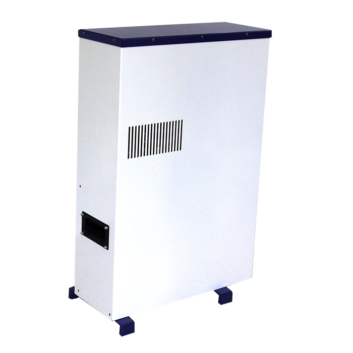
- Supply device;
- Dual focus X-ray emitter;
- Workstation of a laboratory assistant with a high-resolution monitor 24 ″;
- Workstation of a doctor with a high-resolution monitor of a doctor 24 “;
- Medical film printer;
- A set of furniture for the workstation of a doctor and an X-ray laboratory assistant; Remote Control;
- Office printer for printing descriptions;
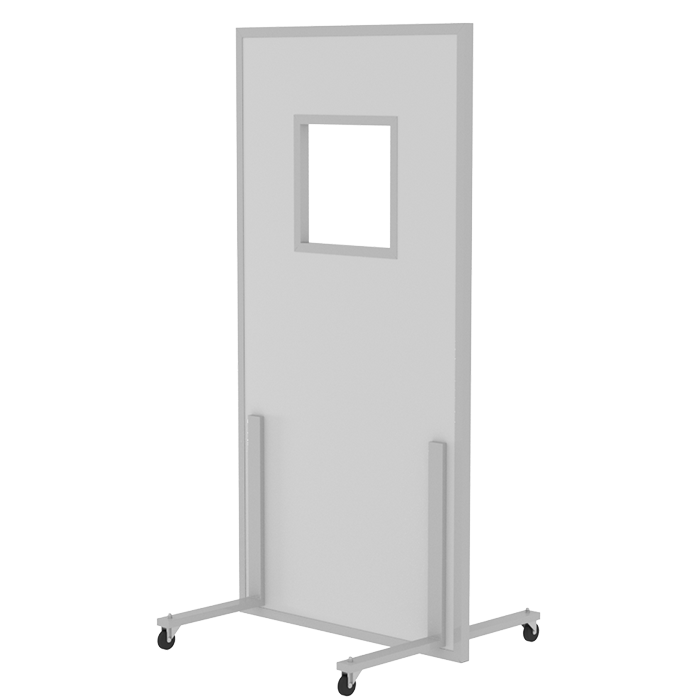
- A set of radiation protection equipment;
- Intercom;
- Dosimeter.
Additional options:
The standard pack can be expanded with various options:
- Vertical shot rack;
- Digital flat panel detector for vertical stand of pictures;
- Additional monochrome diagnostic monitor for the doctor’s workstation;
- Additional workstation;
- PACS system;
- Voltage regulator;
- Additional means of X-ray protection, etc;
- Video surveillance system with two-way audio communication.
The delivery includes: installation, start-up and adjustment works, instruction of the hospital staff by the supplier’s specialists;
Warranty period: 12 months;
Delivery time: within 60 days.
Time of putting the device into operation: within 10 days after delivery, subject to the readiness of the premises for the start of installation work and the presence of an approved technological design.


